
Epidermodysplasia Verruciformis : Microscopic


Comments:
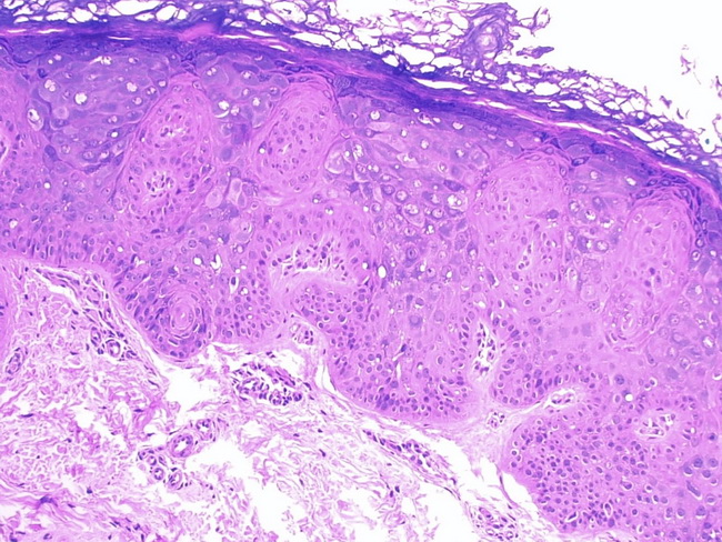
Microscopic Features of Epidermodysplasia Verruciformis: Skin biopsies from the affected areas show acanthosis, hyperkeratosis, and hypergranulosis. The upper epidermis shows large keratinocytes arranged in clusters or columns. There is perinuclear vacuolization, keratohyaline granules, and a striking blue-gray cytoplasm. Advanced lesions show dysplastic changes consisting of nuclear enlargement and hyperchromasia and disorderly maturation (nicely shown in this image). The cytologic atypia extends to appendageal epithelium like sweat ducts. The changes progress to in-situ carcinoma and eventually to invasive carcinoma (30-50% of cases). Image courtesy of: Jose Candido, MD, PhD; Instituto de Patologia de Aracatuba-SP, faculdade de medicina Unisalesiano Aracatuba-SP / Brasil; used with permission.