
Pleomorphic Adenoma : Differential Diagnosis


Comments:
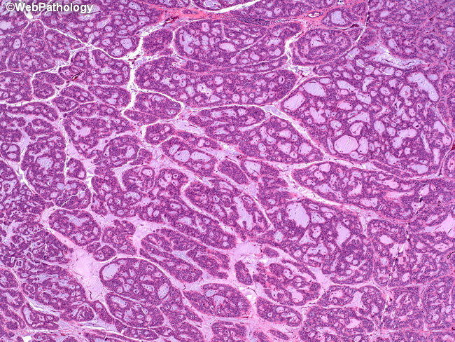
The diagnosis of pleomorphic adenomas is usually straightforward. Diagnostic difficulties may be encountered in cellular lesions located in the minor salivary glands having multiple growth patterns. In addition, due to overlapping histologic features, the distinction between benign and malignant salivary gland tumors may not always be possible in limited biopsies. In such cases, complete excision is recommended. The differential diagnosis includes adenoid cystic carcinoma, monomorphic adenoma, and polymorphous low grade adenocarcinoma. This image depicts cribriform growth pattern in a pleomorphic adenoma that is reminiscent of adenoid cystic carcinoma. The cribriform areas are usually focal and the remainder of the tumor has more typical morphology. Immunohistochemically, adenoid cystic carcinoma shows high Ki-67 index, increased p53 and bcl-2 staining; in contrast, pleomorphic adenomas usually have low Ki-67 index, rare p53 activity and weak bcl-2 staining.