
Malignant Mesothelioma

slide 36 of 48


Comments:
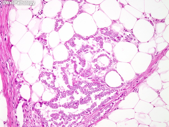
The atypical mesothelial cells line individual fat globules creating a glandular appearance. The presence of linear array of atypical mesothelial cells on free peritoneal or omental surface OR the presence of mesothelial cells in granulation tissue are not necessarily signs of malignancy. They are more likely to be seen in reactive conditions. On the other hand, infiltration of adipose tissue or visceral walls by solid clusters of atypical mesothelial cells is more likely to be encountered in mesotheliomas. This case had clearly diagnostic areas of mesothelioma elsewhere in the specimen.
slide 36 of 48