
Spermatocele : Ultrasound

slide 30 of 61


Comments:
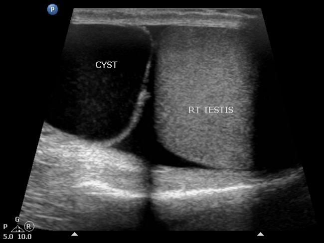
At ultrasonographic examination, spermatoceles are sharply-defined, hypoechoic lesions located near the head of epididymis. They may show fine low-level internal echoes and sometimes have septations. Case courtesy of Dr Rupesh Namdev, Radiopaedia.org. From the case rID: 27587
slide 30 of 61