
Rhabdoid Tumor : Introduction


Comments:
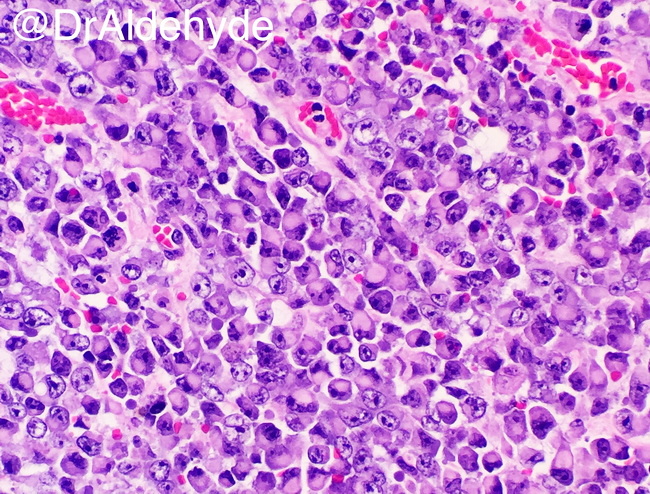
Introduction: Rhabdoid tumor is a rare, highly aggressive malignancy of uncertain histogenesis that belongs to the group of INI1 (SMARCB1)-deficient neoplasms. It is primarily seen in infants and children, although rare adult cases have been described. The tumor typically involves kidney and brain (atypical teratoid/rhabdoid tumor), although extrarenal and extra-CNS tumors have been reported in soft tissues and many other organs/sites. Note: Focal rhabdoid appearance can be seen in other renal neoplasms, including mesoblastic nephroma, Wilms tumor, and renal cell carcinoma. In addition, "rhabdoid phenotype" may develop in a disparate group of neoplasms, including synovial sarcoma, desmoplastic small round cell tumor, rhabdomyosarcoma, malignant melanoma, and many other types of carcinomas. Microscopically, rhabdoid tumor is composed of loosely cohesive sheets of monomorphous cells with eccentrically placed vesicular nuclei, prominent nucleoli, and intracytoplasmic eosinophilic inclusions. The tumor cells often appear plasmacytoid or rhabdomyosarcomatous. Image courtesy of: Woo Cheal Cho, MD; @DrAldehyde; used with permission.