
Spindle Cell Lipoma : Differential


Comments:
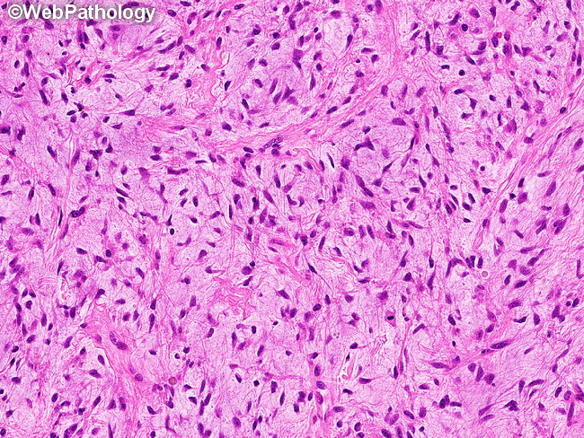
Differential Diagnosis: Spindle cell lipomas are challenging to diagnose specially when they contain little to no fat. This image shows a fat-free spindle cell lipoma with prominent myxoid matrix resulting in a close resemblance to a myxoma. The differential diagnosis of spindle cell lipoma includes:Dermatofibrosarcoma protuberans (location in dermis; prominent storiform pattern with infiltrative growth; younger patient; absence of collagen bundles)Nodular fasciitis (heterogenous appearance; myofibroblastic cells with tissue culture appearance; positive for SMA; lack of CD34 staining)Angiomyofibroblastoma (genital location; prominent vasculature consisting of thick-walled blood vessels; epithelioid cells)Solitary Fibrous Tumor (positive for STAT6 by immunohistochemistry; lacks 13q14 deletion)Cellular angiofibroma and mammary-type myofibroblastoma. Both entities share morphologic and cytogenetic features (deletion of 13q14) and the distinction from spindle cell lipoma may not be possible nor clinically relevant. (differential diagnosis continues in the next slide).