
Pulmonary LCH : Imaging
Home
 Hematopathology
Myeloid, Histiocytic & Dendritic Cell Neoplasms
Langerhans Cell Histiocytosis
Pulmonary LCH : Imaging
Hematopathology
Myeloid, Histiocytic & Dendritic Cell Neoplasms
Langerhans Cell Histiocytosis
Pulmonary LCH : Imaging

slide 23 of 69


Comments:
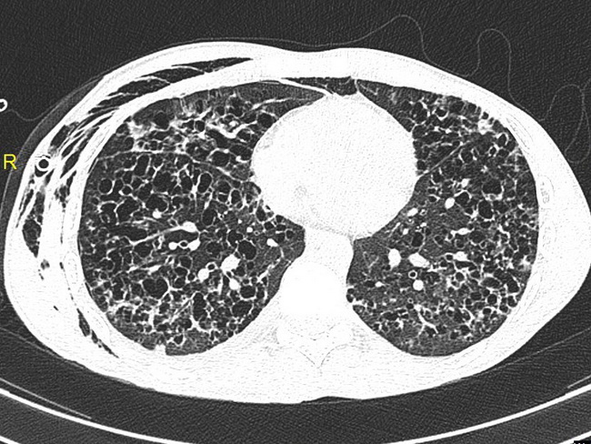
Pulmonary Langerhans Cell Histiocytosis - Diagnosis: Imaging studies play an important role in the diagnosis of pulmonary LCH. Plain X-ray films show bilateral diffuse cystic changes and reticulonodular infiltrates in the lungs with predilection for upper lobes (see previous image). High resolution chest CT (with contrast) will show small symmetric nodules, interstitial fibrosis, cyst formation and honeycombing as seen in this image. The diagnosis can be made on transbronchial biopsy or bronchoalveolar lavage (BAL). BAL requires >5% CD1a+ or CD207+ cells in the appropriate clinical context and imaging findings. Case courtesy of Dr Kashif Nadeem Liaqat, Radiopaedia.org. From the case rID: 29900
slide 23 of 69