
Malakoplakia of Bladder

slide 23 of 36


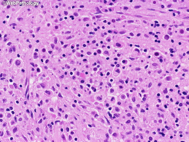
Comments:
Malakoplakia of the urinary bladder. High power view shows abundant foamy histiocytes (von Hansemann cells) and basophilic targetoid structures called Michaelis-Gutmann bodies. These laminated targetoid structures are formed by calcium phosphate deposition on bacteria or their fragments and can be highlighted by calcium stains.
slide 23 of 36