
Chondroblastic Osteosarcoma

slide 20 of 93


Comments:
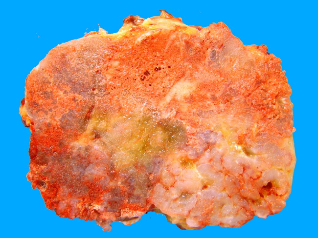
Chondroblastic osteosarcoma involving proximal tibia in a 15 year old male. Bluish-white areas of cartilaginous differentiation are scattered throughout the tumor. Microscopically, these tumors show malignant-appearing chondrocytes in lacunae, crowded areas of spindle cells at the periphery of the lobules, and islands of feathery osteoid matrix within the cartilaginous areas. About 20% of osteosarcomas are classified as chondroblastic. Courtesy of : Dr. Chandra Krishnan. Used with permission.
slide 20 of 93