
Mucinous Cystic Neoplasm : Radiology


Comments:
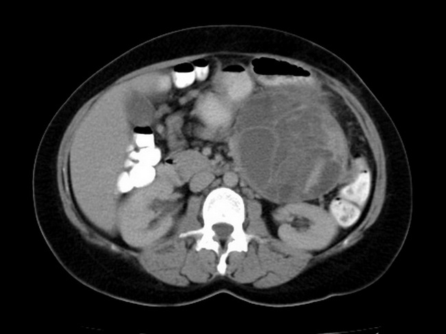
Radiologic Features of Mucinous Cystic Neoplasm (MCN) of Pancreas: On abdominal CT, it appears as a large well-defined thick-walled multilocular cyst that does not communicate with the pancreatic duct system, which is of normal caliber. It has internal septations as well as peripheral "egg-shell" calcifications (20-25% of cases). MCNs with an associated invasive carcinoma are often large, show irregular thickening of septations, and intracystic mural nodules or adjacent solid mass. Case History: 55 y/o female who presented with dyspepsia, epigastric pain and a palpable abdominal mass. Abdominal CT with contrast shows a large multilocular cysitc lesion arising from the pancreatic tail. It has multiple cystic areas with enhanced septae, contents of variable density and foci of calcification within the wall. It displaces the surrounding bowel loops. The diagnosis of mucinous cystic neoplasm was confirmed histologically. Case courtesy of Dr Ahmed Abdrabou, Radiopaedia.org. From the case rID: 26352