
Aneurysmal Bone Cyst : Case 8


Comments:
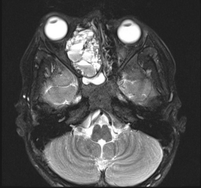
Case History: This is a case of aneurysmal bone cyst (ABC)-like changes arising in association with a psammomatoid juvenile ossifying fibroma (PJOF) of ethmoid in a 10 y/o male. The patient presented with right-sided nasal blockage and facial pain. CT showed a large expansile bone-based mass with a thin bony rim, centered in the right ethmoidal sinuses. MRI revealed a large, fluid-filled mass with a solid rim, compressing on the medial orbital wall but without breaching it. Multiple fluid-fluid levels seen in this MRI axial T2 fat sat. image are highly suggestive of an ABC. Histology: Sections revealed PJOF composed of cellular fibrous tissue with mineralized component and small ossicles resembling psammoma bodies. In addition, there were pseudocystic spaces filled with blood and lined by fibroblastic tissue, hemosiderin-laden macrophages, and osteoclast-type giant cells. The morphologic features were diagnostic of ABC-like changes in association with PJOF.