
Solid Pseudopapillary Tumor

slide 13 of 32


Comments:
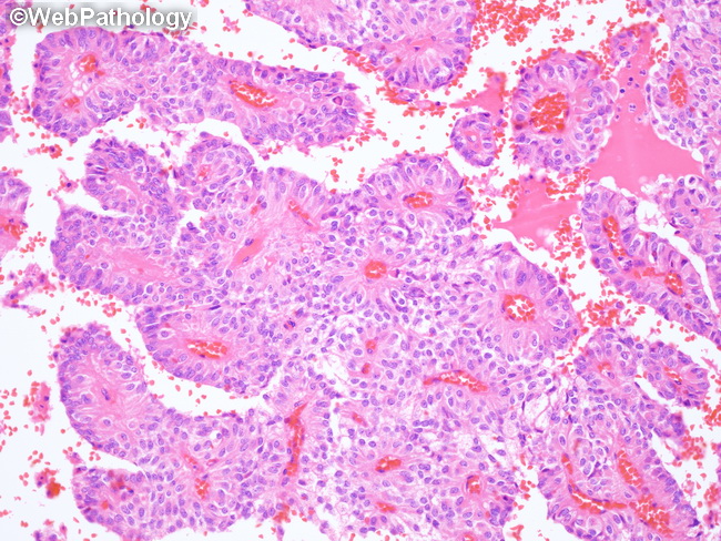
Microscopic Features of Solid Pseudopapillary Tumor (SPT) of Pancreas (continued): The pseudopapillary structures are formed due to degenerative changes and cellular discohesion which leaves 1 to 2 layers of tumor cells around hyalinized fibrovascular cores. In cases with advanced degenerative changes, there are scattered foci of cystic change (or even large cysts), hemorrhage, microcalcifications (or ossification), cholesterol crystals, multinucleated giant cells, and aggregates of foamy histiocytes. PAS-positive intra- and extracytoplasmic eosinophilic hyaline globules may be present. Foci of myxoid change around blood vessels may be seen.
slide 13 of 32