
Atypical Adenomatous Hyperplasia : Lung

slide 116 of 128


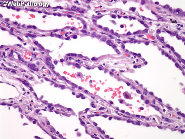
Comments:
Higher magnification of atypical adenomatous hyperplasia showing proliferation of mild to moderately atypical type II pneumocytes along alveolar surfaces. Differential Diagnosis : Adenocarcinoma-in-situ (AIS): The spectrum of histologic changes between AAH and AIS is a morphologic continuum and distinction between the two can be difficult. AIS is usually larger than 0.5 cm, shows cellular crowding, and more abrupt transition to adjacent normal alveolar lining cells than in AAH. Reactive Pneumocyte Hyperplasia: This lesion is usually seen in the setting of parenchymal inflammation or fibrosis. AAH is rarely accompanied by interstitial fibrosis or inflammation.
slide 116 of 128