
Extracavitary Primary Effusion Lymphoma


Comments:
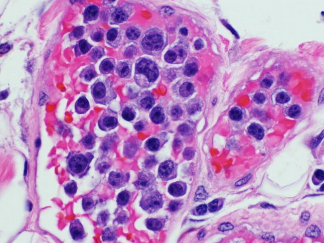
Extracavitary Primary Effusion Lymphoma (PEL): Lymphomas resembling PEL may rarely develop as solid masses in the absence of an effusion. They involve lymph nodes or extranodal sites such as gastrointestinal tract. Most cases are similar to PEL in all aspects, including clinical presentation, association with HIV, HHV8, and EBV, morphology, immunophenotype, and clonal immunoglobulin gene rearrangements. However, in 25% of cases, these solid HHV8-positive lymphomas lack CD45 (PEL is CD45+) and express B-cell markers CD20, CD79a, and CD138 (PEL is negative for these markers). In addition, there may be aberrant expression of T-cell markers (just as in PEL). Morphologically, the extracavitary PEL can resemble anaplastic large cell lymphoma (ALCL) or immunoblastic/plasmablastic lymphomas. Some cases show prominent sinusoidal infiltration mimicking metastatic carcinoma. Immunostaining for HHV8 is helpful in distinguishing them from ALCL. Large pleomorphic cells resembling Reed-Sternberg or Hodgkin cells may be present. Some cases show invasion of endothelial-lined lymphatic or vascular spaces and resemble intravascular lymphoma. At genetic level, they lack abnormalities in MYC, BCL2, BCL6, and CCND1. They are positive for both EBV and HHV8. The image shows primary effusion lymphoma which presented initially as intravascular lymphoma confined to dermal vessels. Subsequently, the patient developed pleural effusion. The patient was immunosuppressed and the lymphoma was HHV8-positive and EBV-positive. Image courtesy of: Genevieve Crane, MD, PhD.