
Amyloid Lymphadenopathy


Comments:
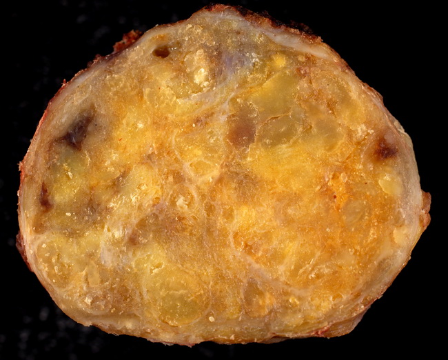
Introduction: Proteinaceous lymphadenopathy refers to deposition of amorphous proteinaceous amyloid-like material in lymph node with partial or total effacement of the nodal architecture. In primary or secondary amyloidosis, lymph nodes are involved in 17% to 37% of cases. Clinical Features: More than 90% of patients with primary amyloidosis have a monoclonal M protein in urine, serum, or cerebrospinal fluid. Multiple myeloma is seen in 20% of patients with AL amyloidosis. If AL protein is detected in amyloid deposits in a lymph node, further work up is warranted to rule out plasma cell dyscrasia, idiopathic amyloidosis, or a lymphoproliferative neoplasm. Work up includes bone marrow examination and protein electrophoresis on serum and urine specimens. Secondary amyloidosis (with deposition of amyloid fibril protein AA) can be seen with a number of neoplastic and non-neoplastic conditions with high cell turnover, including rheumatoid arthritis, ankylosing spondylitis, Crohn disease, tuberculosis, and osteomyelitis. About 20% of uremic patients with lymphadenopathy have AA amyloid deposits in the lymph nodes. Case History: The patient was a 54 y/o male who presented with isolated inguinal lymphadenopathy without any systemic involvement (a rare presentation). The lymph node measured 4.5 cm and was firm with a yellow-brown waxy surface on cutting. The diagnosis of amyloid lymphadenopathy was confirmed with special stains (next few slides). Image courtesy of : Ed Uthman, MD, Houston, Texas. Used with permission.