
Polymorphous Low-grade AdenoCA
Home
 Head & Neck
Salivary Glands
Malignant Neoplasms of Salivary Glands - II
Polymorphous Low-grade AdenoCA
Head & Neck
Salivary Glands
Malignant Neoplasms of Salivary Glands - II
Polymorphous Low-grade AdenoCA

slide 15 of 21


Comments:
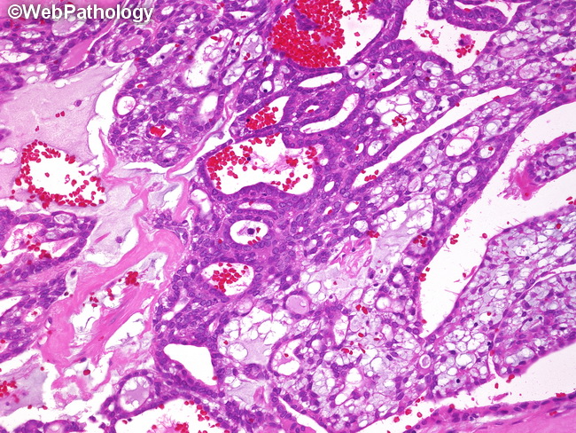
The image shows polymorphous low-grade adenocarcinoma (PLGA) composed of tubulo-glandular structures in a background of myxoid stroma. In some cases, the stroma may be hyalinized or even densely sclerotic (see next image). Immunohistochemical profile: PLGA is positive for cytokeratins, EMA, and S-100 protein. There is variable but at least focal positivity for p63, whereas p40 is usually negative. A p63/p40 panel is sometimes used in distinguishing PLGA (p63+/p40-) from pleomorphic adenoma (p63+/p40+) and adenoid cystic carcinoma (p63+/p40+). Ki-67 proliferation index is low. Immunohistochemistry is usually not essential in making the diagnosis of PLGA.
slide 15 of 21