
Condyloma Acuminata : Microscopic

slide 20 of 36


Comments:
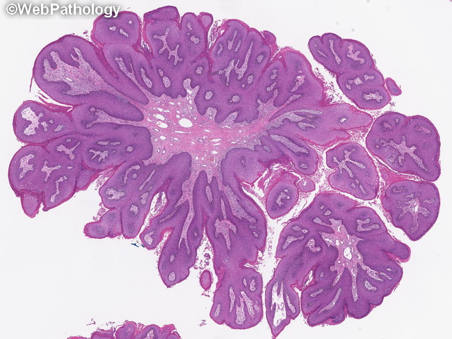
Genital condylomas show marked acanthosis, papillomatosis, hyperkeratosis, and parakeratosis. Superficial layers show vacuolated keratinocytes (koilocytes) and prominent coarse keratohyalin granules. Binucleated cells, dyskeratocytes, and wrinkled nuclei (resinoid appearance) may also be seen. Cytologic atypia is minimal and mitotic figures are confined to the basal region.
slide 20 of 36