
Systemic Mastocytosis : Bone Marrow


Comments:
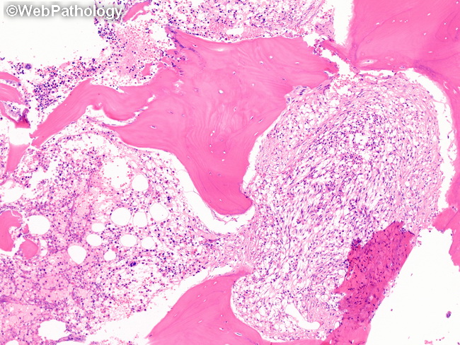
Bone Marrow Involvement in Systemic Mastocytosis (SM): The most common finding in bone marrow is multifocal or disseminated, paratrabecular and perivascular infiltrates of mixed cellularity, with a granulomatous appearance. Round, oval, or spindle-shaped mast cells are present within these infiltrates and can be recognized with the help of immunostains ( positive for tryptase and CD25). Other cell types include eosinophils, plasma cells, histiocytes, and fibroblasts. A dense network of reticulin fibers is frequently present. Collagen fibrosis develops in long-standing cases of SM. Clinical History: This bone marrow biopsy is from an adult female with indolent systemic mastocytosis (ISM). The diagnosis of ISM requires at least one major criterion and one minor criterion or at least 3 minor SM criteria and absence of features of more advanced types of SM. The right half shows replacement of marrow by a compact spindle cell infiltrate that was strongly positive for tryptase and CD117 confirming them to be mast cells. The left half shows an area of hematopoiesis with reduced number of precursors. The patient had previous diagnosis of urticaria pigmentosa. Her peripheral blood smear was normal. There was no organomegaly.