
Systemic Mastocytosis : Bone Marrow


Comments:
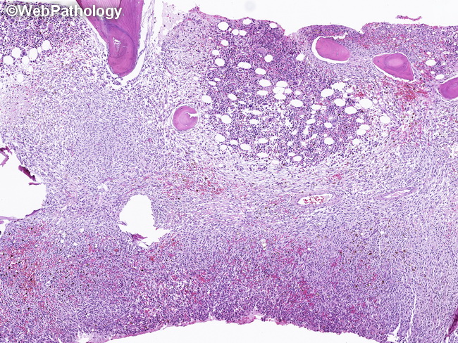
Bone Marrow Involvement in Systemic Mastocytosis (SM): Bone marrow is the most frequently involved extracutaneous site in SM. Bone marrow aspirate and trephine biopsy from iliac crest are required for the diagnosis, staging, and grading of SM. An immunohistochemical panel consisting of tryptase, CD117/KIT, CD25, and CD2 is useful in detecting mast cell (MC) infiltrates and assessing their pattern of infiltration. Normally, MC constitute <0.1% of all the nucleated cells in the bone marrow and do not express CD25 or CD2. Depending upon the subtype of SM, the mast cell types found in bone marrow aspirates can include: immature MC (metachromatic blast), atypical MC type I (hypogranulation, spindling, and oval/eccentric nuclei), atypical MC type II (promastocyte; bilobed or multilobed nuclei), and mature tissue MC. This image shows diffuse, compact MC infiltrate with effacement of normal marrow architecture. There is loss of marrow fat and only isolated pockets of hematopoiesis are present (seen here along the upper edge of the image). This pattern is usually seen in mast cell leukemia, and some cases of smoldering systemic mastocytosis and aggressive systemic mastocytosis. The patient shows clinical signs of bone marrow failure.