
Urticaria Pigmentosa : Differential


Comments:
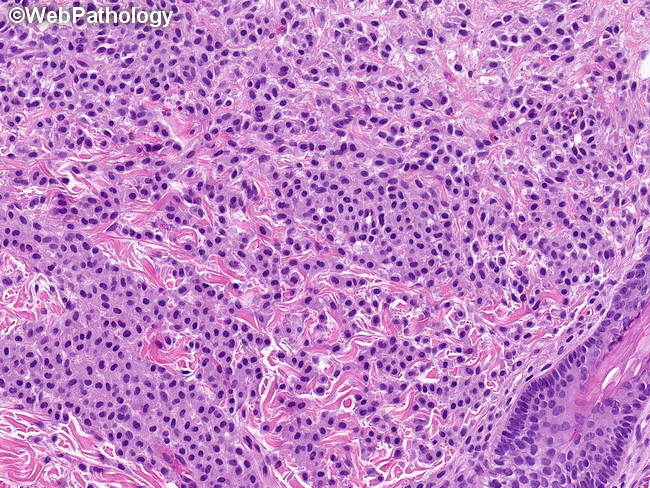
The neoplastic mast cells in mastocytosis are histologically indistinguishable from normal mast cells. They are oval, polygonal, or spindle shaped and have moderate amounts of eosinophilic or amphophilic cytoplasm. Faint cytoplasmic granules may be visible. The nuclei are uniform round or oval with clumped chromatin and inconspicuous nucleoli. Reactive mast cell hyperplasia: Increased number of mast cells may be seen in inflammatory skin disorders. They may overlap with cases of cutaneous mastocytosis in which the mast cell infiltrate is sparse. In reactive conditions, the mast cells are loosely scattered in the dermis, often in a perivascular distribution. There are no compact clusters or dense aggregates and there is no aberrant expression of CD25 and/or CD2 in reactive conditions. The image shows sheets of mast cells in a skin biopsy from a case of urticaria pigmentosa.