
LCH : Morphology
Home
 Hematopathology
Myeloid, Histiocytic & Dendritic Cell Neoplasms
Langerhans Cell Histiocytosis
LCH : Morphology
Hematopathology
Myeloid, Histiocytic & Dendritic Cell Neoplasms
Langerhans Cell Histiocytosis
LCH : Morphology

slide 9 of 69


Comments:
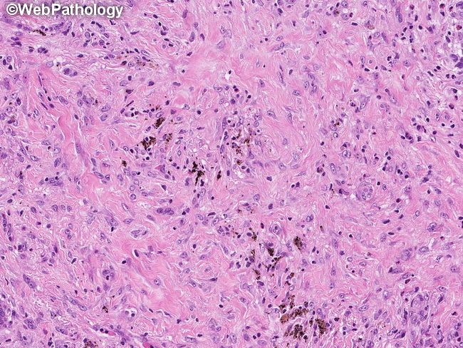
Early lesions of Langerhans cell histiocytosis (LCH) are cellular and show numerous Langerhans cells and eosinophils with minimal or no fibrosis. The later stages are dominated by fibrosis (as shown here) and foamy macrophages with decreased numbers of LC cells and eosinophils. If Langerhans cells display marked pleomorphism, anaplastic features, and brisk mitotic activity with atypical mitoses, the diagnosis of Langerhans cell sarcoma should be considered.
slide 9 of 69