
Histiocytic Sarcoma : Differential


Comments:
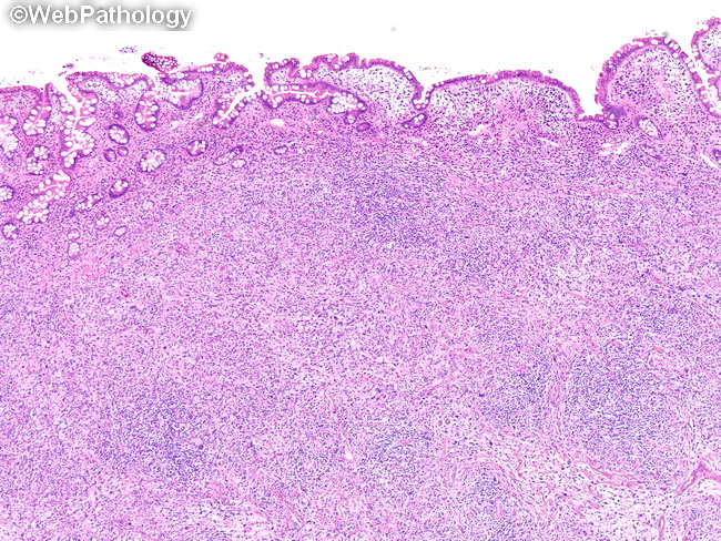
Differential Diagnosis of Histiocytic Sarcoma: Malignant entities: Anaplastic large cell lymphoma, B- or T-lineage large cell lymphomas, hepatosplenic T-cell lymphoma, follicular dendritic neoplasms, myeloid sarcoma (especially those with monoblastic differentiation), anaplastic carcinomas, and malignant melanoma. Benign histiocytic entities: Infection associated hemophagocytic syndrome, familial hemophagocytic lymphohistiocytosis, storage diseases (Gaucher's and Niemann-Pick). Both reactive histiocytic proliferations and histiocytic sarcoma can have hemophagocytosis, multinucleated cells, and foamy cytoplasm. Histiocytic sarcoma shows cytologically malignant nuclei in contrast to bland nuclear features of reactive histiocytic proliferations. Interdigitating dendritic cell sarcoma shows histologic overlap with histiocytic sarcoma. Features favoring interdigitating dendritic cell sarcoma include relatively lower degree of nuclear pleomorphism, greater degree of cell spindling and stronger, diffuse positivity for S-100. Anaplastic large cell lymphoma is the non-Hodgkin lymphoma most frequently confused with histiocytic sarcoma. The distinction of non-Hodgkin lymphomas from histiocytic sarcoma requires immunophenotypic analysis (by flow cytometry) and gene rearrangement studies. The presence of CD30 expression or expression of lineage specific T- or B-cell markers rules out histiocytic sarcoma. The image shows histiocytic sarcoma of small bowel that caused intestinal obstruction. The patient presented to the emergency room with severe abdominal pain, nausea and vomitting. There was history of fever and progessive weight loss. The intestinal architecture is distorted by sheets of pleomorphic tumor cells with abundant eosinophilic cytoplasm. There is an infiltrate of eosinophils, lymphocytes, and plasma cells in the background (images 10-15).