
Mycosis Fungoides : Patch Stage
Home
 Hematopathology
Mature T-cell & NK-cell Neoplasms
Mycosis Fungoides & Sezary Syndrome
Mycosis Fungoides : Patch Stage
Hematopathology
Mature T-cell & NK-cell Neoplasms
Mycosis Fungoides & Sezary Syndrome
Mycosis Fungoides : Patch Stage

slide 19 of 73


Comments:
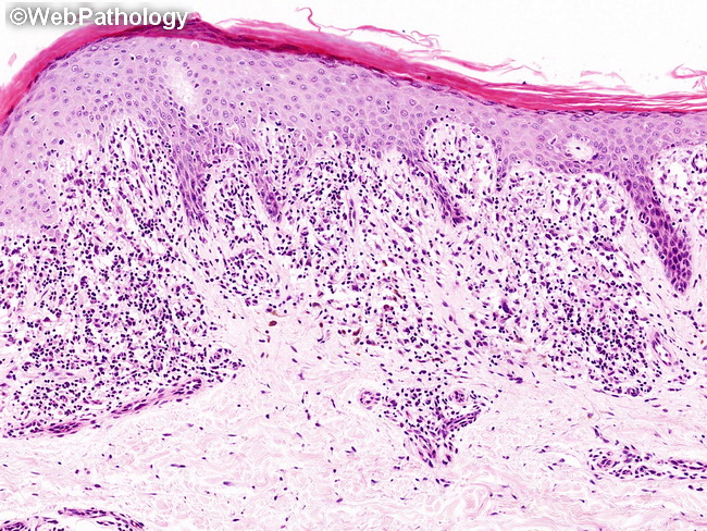
Mycosis Fungoides - Patch Stage: As patches develop, the lymphocytic infiltrate becomes dense and band-like and begins to show atypical lymphocytes with hyperchromatic, irregular, cerebriform nuclei. The atypical lymphoid cells surrounded by small clear haloes may be seen within the epidermis. There is fibroplasia of papillary dermis. Eosinophils, plasma cells, and extravasated red blood cells may be present in small numbers. Pigment incontinence is a common feature. The epidermis can be of normal thickness, acanthotic, or atrophic. There is usually mild hyperkeratosis and focal parakeratosis.
slide 19 of 73