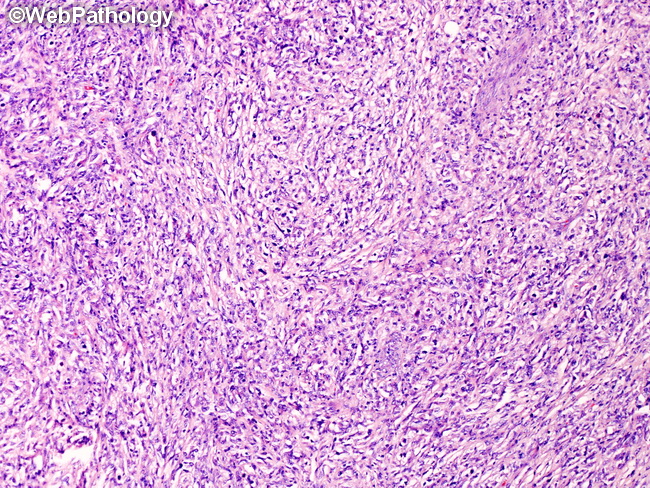
PMLBCL : Diagnosis


Comments:
Diagnosis of Primary Mediastinal Large B-cell Lymphoma (PMLBCL): The location of tumor in the anterosuperior mediastinum necessitates invasive procedures such as mediastinoscopy or percutaneous CT-guided core needle biopsies to get the diagnostic material. The diagnosis is made on the basis of morphologic features in the biopsy specimen, in conjunction with immunohistochemical profile and appropriate clinical presentation. After diagnosis has been established, the next step of clinical staging requires physical examination, whole body CT or PET/CT imaging, bone marrow biopsy, whole blood count, and serum biochemistries. Serum lactate dehydrogenase is elevated in 70-80% of cases. β2-microglobulin is normal. Most patients present in clinical stage I or II. Non-mediastinal PMLBCL: Mediastinal involvement is considered essential for the diagnosis of PMLBCL. However, cases of diffuse large B-cell lymphoma with features of PMLBCL but without detectable mediastinal involvement have been reported. These extra-mediastinal tumors have shown gene expression profile similar to PMLBCL, including alterations in CIITA and PDL1/2 genes.


