
Lymphoepithelial Sialadenitis & MALT Lymphoma


Comments:
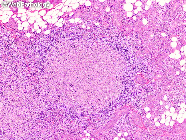
Lymphoepithelial Sialadenitis (LESA) & Salivary Gland MALT Lymphoma: Immunophenotypically, the germinal centers seen in LESA are similar to those in Peyer's patches and lymph nodes. They are surrounded by polyclonal CD20+, IgM+, and IgD+ mantle zone cells. The interfollicular areas are occupied by CD3+ T-cells which may sometimes outnumber CD20+ B-cells. Polytypic plasma cells are also usually present. This image of LESA shows a germinal center with well-defined mantle zone. The adjoining salivary gland parenchyma is atrophic with predominantly small ducts, a few small acini and fatty infiltration. The histologic features of LESA and marginal zone lymphoma form a spectrum and there are no sharp distinctions. Some cases of LESA show monoclonal B-cell clones with immunoglobulin gene rearrangement studies. Distinction between LESA and MALT lymphoma can be difficult at times.