
Lymphoepithelial Sialadenitis & MALT Lymphoma
Home
 Hematopathology
Mature B-cell Neoplasms - Part I
Extranodal Marginal Zone Lymphoma
Lymphoepithelial Sialadenitis & MALT Lymphoma
Hematopathology
Mature B-cell Neoplasms - Part I
Extranodal Marginal Zone Lymphoma
Lymphoepithelial Sialadenitis & MALT Lymphoma

slide 14 of 100


Comments:
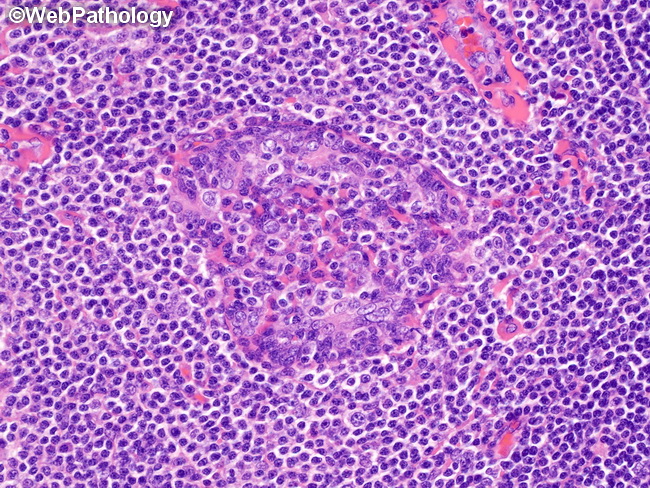
Lymphoepithelial Sialadenitis (LESA) & Salivary Gland MALT Lymphoma: In the later stages of LESA, the lymphoepithelial lesions show partial or complete occlusion of ductal lumens. There is atrophy of columnar ductal epithelium, proliferation of basal epithelial cells and infiltration by marginal zone B-cells. In some cases, the lymphoepithelial structures undergo extensive cystic degeneration forming a multicystic gland. This image shows a lymphoepithelial lesion surrounded by marginal zone B-cells with pale staining cytoplasm.
slide 14 of 100