
Lymphoepithelial Sialadenitis & MALT Lymphoma


Comments:
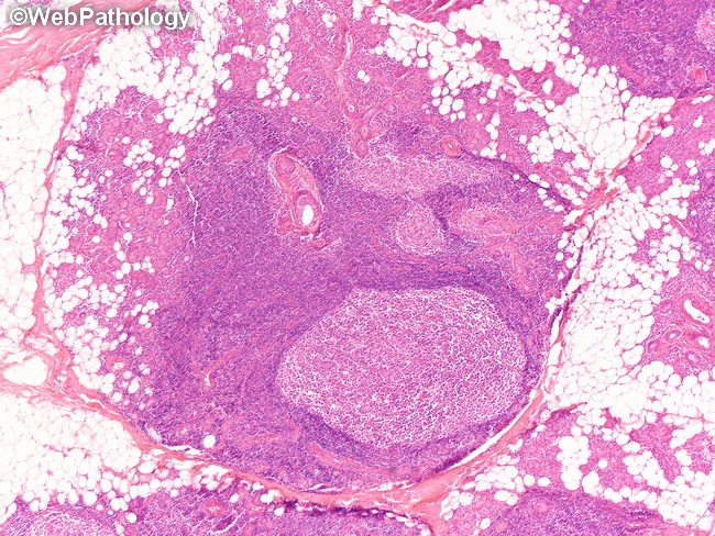
Lymphoepithelial Sialadenitis (LESA) & Salivary Gland MALT Lymphoma: Normal salivary glands (especially parotid) may contain lymph nodes but no organized lymphoid tissue. In the setting of chronic inflammation of various etiologies, salivary glands can acquire lymphoid tissue resembling Peyer's patches. The prototypic lesion is lymphoepithelial sialadenitis (LESA) in patients with Sjogren syndrome; however, similar appearance can be seen in other inflammatory or autoimmune conditions or even in the absence of a known etiology. In LESA, there is lymphoid infiltration of salivary gland tissue with atrophy and destruction of acini and formation of lymphoepithelial lesions. LESA lesions respond to corticosteroids, but they can progress to lymphomas. The association between LESA (in the setting of Sjogren syndrome) and development of salivary gland lymphomas is well-known. Sjogren syndrome patients have a 15-fold higher incidence of non-Hodgkin lymphomas, especially extranodal marginal zone lymphomas. This image of LESA shows lymphoid follicles surrounding salivary ducts, acinar atrophy, and fatty replacement of parenchyma.