
Helicobacter Pylori Gastritis & MALT Lymphoma


Comments:
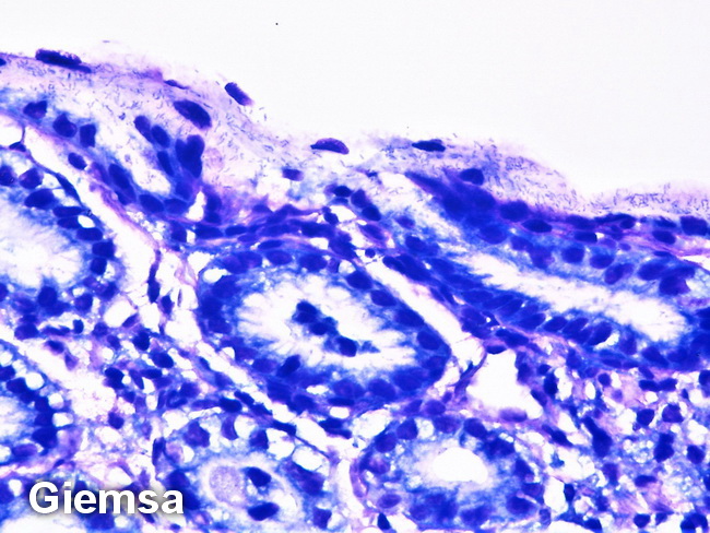
Helicobacter pylori Gastritis & Gastric MALT Lymphoma: H. pylori infection results in active chronic inflammation with formation of B-cell follicles. Marginal zone B-cells infiltrate into adjacent glands forming lymphoepithelium. The interfollicular areas contain T-cells, plasma cells, macrophages, and, sometimes, neutrophils. H. pylori are slightly curved, flagellated gram-negative rods that are usually seen in gastric mucus, over foveolar epithelium, and gastric pits. When they are numerous, they can be easily seen with H&E stain, but special stains like Giemsa are helpful in confirming the diagnosis (as shown here). In cases of H. pylori gastritis with florid lymphoid hyperplasia (FLH), the distinction from MALT lymphoma may be difficult but it can be done with immunophenotypic profiling. The germinal centers in FLH are surrounded by IgM+, IgD+ mantle zone cells, where as MALT lymphoma cells are IgM+ and IgD-ve. The presence of immunoglobulin light chain-restricted B-cells and plasma cells supports MALT lymphoma; however, the presence of polyclonal cell populations does not rule out the diagnosis of lymphoma. Image courtesy of: @PatholWalker.