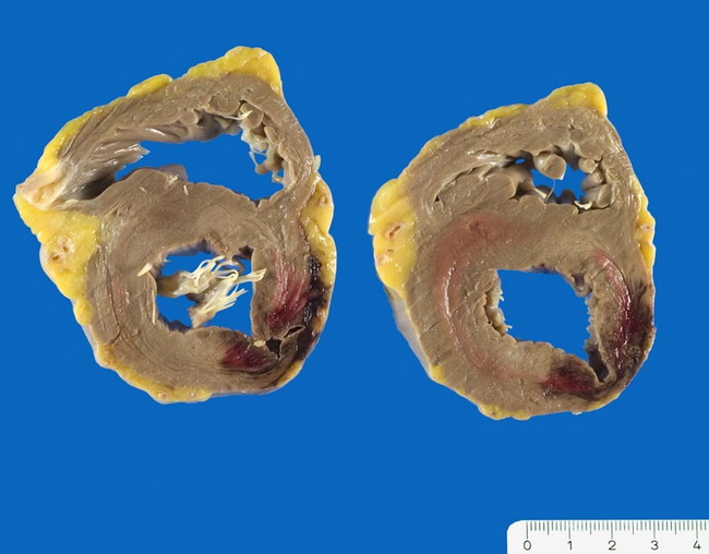
Myocardial Infarction - Rupture of Free Wall


Comments:
Transmural infarction is sometimes complicated by myocardial rupture. The most common form is the rupture of the free left ventricular wall as shown in this image. There was 300 ml blood in the pericardial space (tamponade). The patient was a 67 y/o female with biventricular concentric myocardial hypertrophy and severe coronary artery disease. Rupture of the interventricular septum is less common. The least common form of myocardial rupture following acute myocardial infarction is the rupture of papillary muscles. Image copyright: pathorama.ch.


