
Adamantinomatous Craniopharyngioma

slide 6 of 32


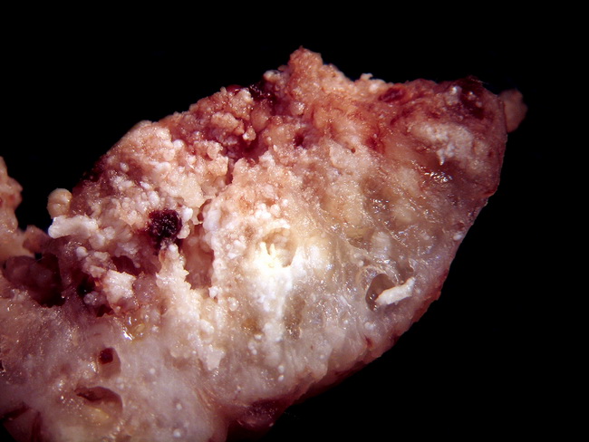
Comments:
Resected specimen of an adamantinomatous craniopharyngioma showing solid and small cystic areas as well as yellowish-white nodules which correspond to nodules of wet keratin seen microscopically. Courtesy of: Dr. Luciano de Souza Queiroz, Dept. of Pathology, Faculty of Medical Sciences, State University of Campinas (UNICAMP), Campinas, Sao Paulo State, BRAZIL. Additional images are here.
slide 6 of 32