
B-cell Prolymphocytic Leukemia : Microscopic


Comments:
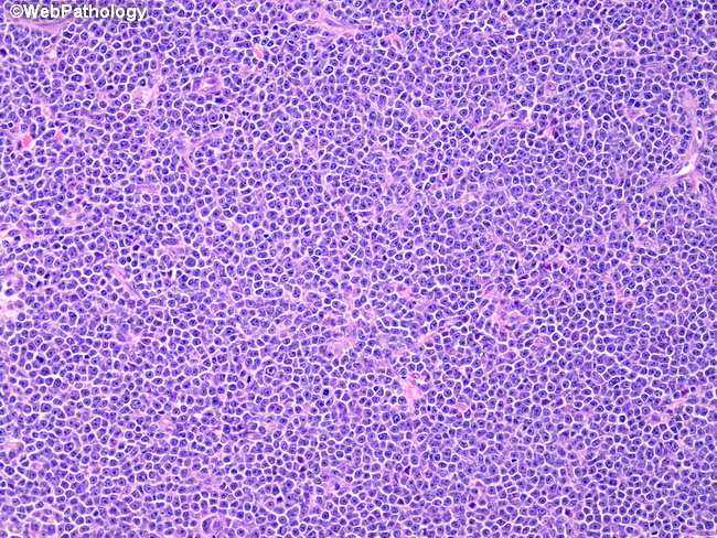
Microscopic Features: The diagnosis of B-cell prolymphocytic leukemia (B-PLL) is typically not made via tissue biopsy but by morphologic, immunophenotypic, and genetic analysis of peripheral blood. However, a tissue biopsy is useful in excluding other more common lymphoid malignancies such as mantle cell lymphoma and splenic marginal zone lymphoma which have overlapping features with B-PLL. The prolymphocytes are uniform, intermediate to large size with moderate amount of cytoplasm, round or oval nucleus, and a prominent nucleolus. The bone marrow infiltrates are interstitial or nodular and show sheets of prolymphocytes. Spleen involved by B-PLL shows expansion of white pulp areas as well as infiltration of red pulp by prolymphocytes. Lymph nodes involved by B-PLL (this image) show diffuse effacement of architecture by prolymphocytes. Unlike CLL, proliferation centers are not seen.