
Apocrine Cystadenoma

slide 61 of 126


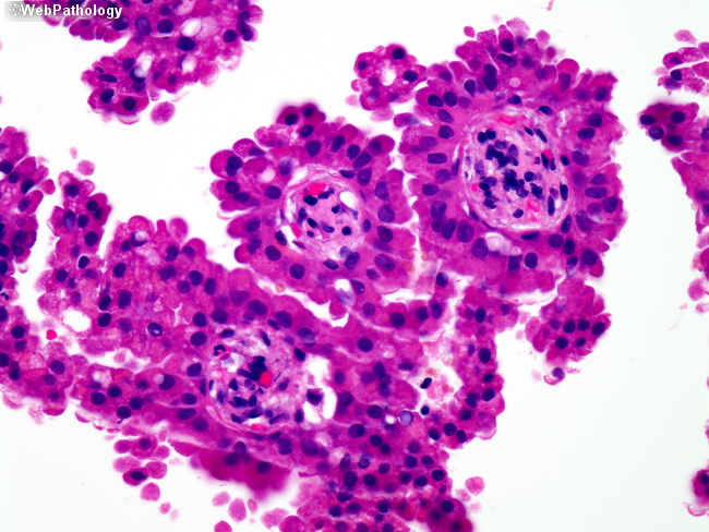
Comments:
The above photomicrograph illustrates a high magnifcation view of apocrine papillae, with true fibrovascular cores and decaptiation secretion. Lesions with extensive papillary formation and/or nuclear atypia should be removed by complete excision.
slide 61 of 126