
Uterine Hemangioma


Comments:
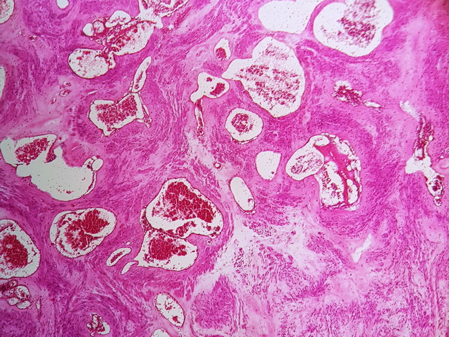
Microscopically, uterine hemangiomas consist of irregularly-shaped cavernous spaces infiltrating between the myometrial fascicles. The vascular spaces are lined by flattened endothelium (positive for CD31 and CD34) and distended with blood. Uterine hemangiomas are classified into congenital (associated with hereditary syndromes) and acquired (linked to high estrogen levels). Given their rarity and non-specific presentation, correct diagnosis is rarely made before surgery. Vaginal examination, uterine curettage, ultrasonography, and hysterography are usually unhelpful. Pelvic angiography, CT scan with contrast, or MRI may confirm vascular nature of the lesion. Most cases are considered to be adenomyosis preoperatively. The initial treatment is conservative and may consist of carbon dioxide laser excision, cryotherapy, radiation, electrocauterization, uterine artery embolization, and laser ablation. Hysterectomy is offered if conservative measures fail. Reference: Chou, W and Chang H. Uterine Hemangioma : A Rare Pathologic Entity. Arch Pathol Lab Med. 2012;136:567-571. Case courtesy of: Vahid Ariabod, MD, Dept. of Pathology, Islamic Azad University, Mashhad, Iran; used with permission.