
Leiomyoma of Uterus : Submucosal

slide 3 of 49


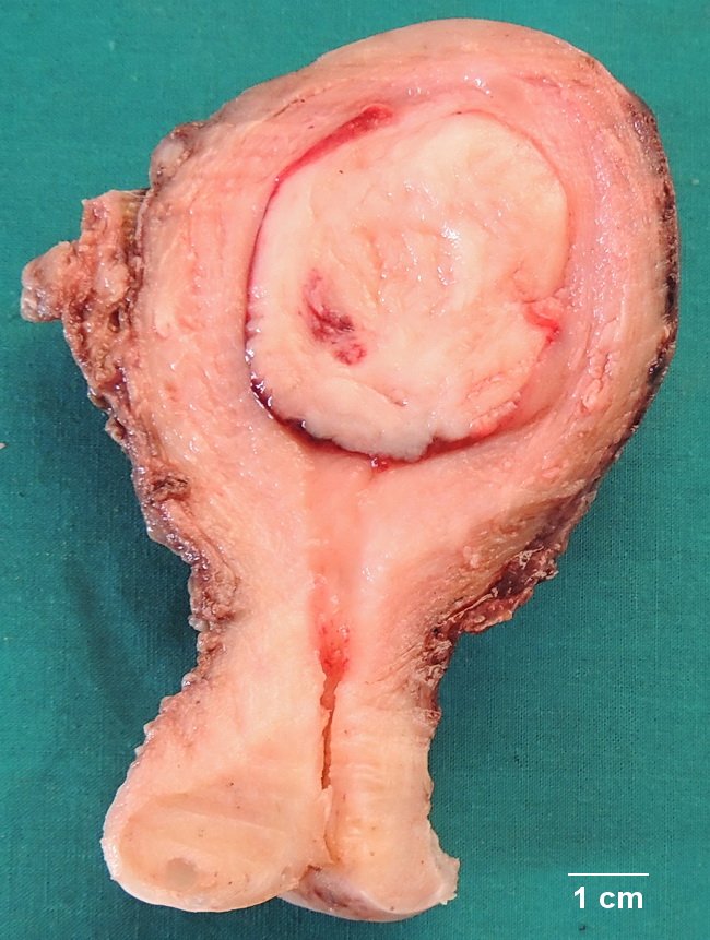
Comments:
This specimen is from a 36 y/o female who presented with abdominal pain and menorrhagia. Sonography revealed a mass in the uterine cavity. The hysterectomy specimen shows a 4 cm submucosal leiomyoma obliterating the endometrial cavity. It was attached to the uterine wall via a thin pedicle. The cut surface has pink-tan whorled appearance. Based on their location, the uterine leiomyomas may be intramural, subserosal, cervical, or submucosal (as in this case). Case courtesy of: Dr. Sanjay D. Deshmukh, Professor of Pathology. Dr. Vithalrao Vikhe Patil Medical College & Hospitals, Ahmednagar, India.
slide 3 of 49