
Ovary : Leiomyosarcoma

slide 5 of 5

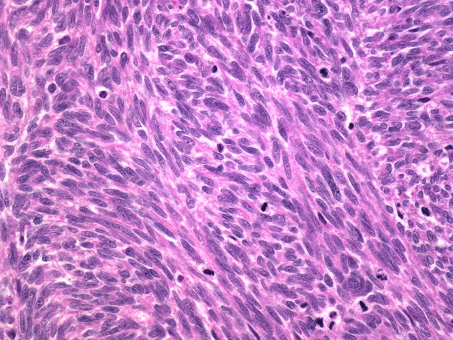
Comments:
High power view of the previous image showing tumor cells in intersecting fascicles. The tumor showed moderate cytologic atypia and >20 mitotic figures/10 hpf. The differential diagnosis of ovarian leiomyosarcomas includes fibrosarcomas, spindle cell carcinomas, and metastatic GIST. Majority of the cases recur following surgery and the overall prognosis is grim.
slide 5 of 5