
Steroid Cell Tumor, NOS : Case Presentation


Comments:
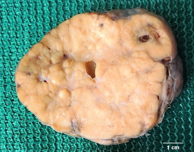
Case History: A 21 y/o obese female presented with amenorrhea and hirsutism (chin, upper lip and chest wall). On examination, she had atrophic breasts. The laboratory findings revealed elevated serum total testosterone level (560 ng/dl; reference range <60 ng/dl) with normal LH, FSH, DHEA, DHEA-S, 24-hr urine cortisol, and 17-hydroxyprogesterone levels. Imaging studies with pelvic ultrasound and CT of abdomen revealed a unilateral solid ovarian tumor without local invasion or metastasis. Based on above findings the tumor was surgically resected. Gross examination showed a well circumscribed, 8 cm neoplasm with thin capsule, and yellow to yellow-brown, lobulated cut surface. Microscopic examination confirmed the diagnosis of Steroid cell tumor, NOS. Case courtesy of: Dr. Sanjay D. Deshmukh and Dr. Baba B. Shinde, (Professors, Dept. of Pathology), Dr. Vithalrao Vikhe Patil Foundation's Medical College & Hospitals, Ahmednagar, India.