
Thecoma : Differential Diagnosis


Comments:
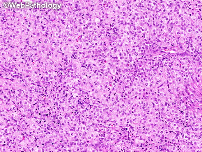
Differential Diagnosis: The differential diagnosis of thecoma (shown here) includes: fibroma, adult granulosa cell tumor, and sclerosing stromal tumor.Fibroma: Fibroma and thecoma have overlapping morphology and the two may coexist within the same tumor (fibrothecoma). The distinction between the two is largely an academic exercise since both are benign. The term thecoma is applied to stromal tumors with large amount of cytoplasmic lipids and/or clinical evidence of steroid hormone production (endometrial hyperplasia, carcinoma). Grossly, thecomas are often yellow rather than white. They are positive for inhibin and CD10 (which may be focal). In contrast, fibromas are non-functioning spindle cell tumors with a collagenous stroma and scant amounts of cytoplasmic lipid. Fibromas are negative (or weakly +ve) for CD10 and show only patchy positivity for inhibin. (continued in the next image).