
Thecoma : Microscopic Features

slide 26 of 74


Comments:
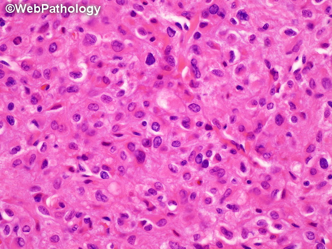
Microscopic Features of Thecoma: (continued) The tumor cells have round to oval nuclei with finely dispersed chromatin, moderate amount of pale vacuolated (lipid-rich) or greyish-pink cytoplasm and indistinct cell borders creating a syncytial appearance. Punctate nucleoli and nuclear grooves are sometimes present. There is no cytologic atypia, although degenerative-type atypia without increased mitotic activity may be present.
slide 26 of 74