
Ovarian Carcinoid : Trabecular

slide 148 of 183


Comments:
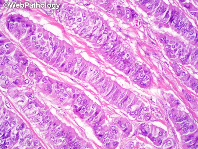
Microscopic Features of Ovarian Carcinoids - Trabecular Pattern: In the trabecular pattern, the neoplastic cells form long parallel, serpentine ribbons, trabeculae and cords that are separated by variable amounts of stroma. The tumor nuclei are elongated and arranged perpendicular to the axis of the trabeculae. Trabecular pattern is similar to the hindgut carcinoids seen in the rectum. It is usually associated with mature cystic teratoma and is often mixed with the insular pattern.
slide 148 of 183