
Ovarian Carcinoid : Microscopic

slide 140 of 183


Comments:
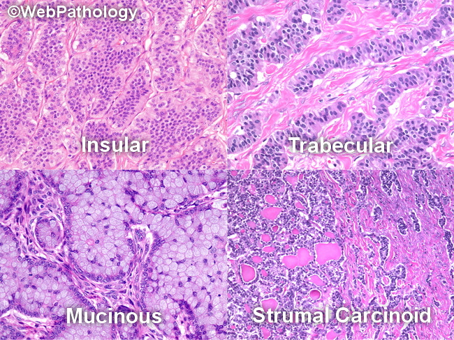
Microscopic Features: The morphology of primary ovarian carcinoids is similar to that seen in other locations. Five architectural patterns have been recognized: insular (50% of cases), strumal carcinoid (40% of cases), trabecular, mucinous, and mixed type. The tumor cells have abundant eosinophilic cytoplasm and may show red-brown argentaffin granules. The nuclei are uniform and have salt and pepper chromatin. Mitotic activity is low. Well-differentiated mucinous glands are seen in about 40% of cases. Teratomatous elements may be found in the ipsilateral or the contralateral ovary. Some ovarian carcinoids are associated with Brenner tumor or mucinous cystadenoma.
slide 140 of 183