
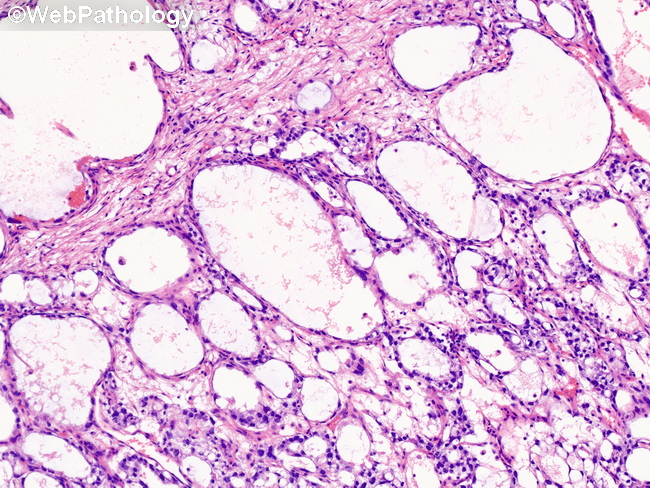
Yolk Sac Tumor : Polyvesicular Vitelline Pattern

slide 116 of 183


Comments:
Polyvesicular Vitelline Pattern: This uncommon pattern (seen in 25% of yolk sac tumors) shows vesicles lined by flattened bland tumor cells surrounded by loose myxoid or fibrous stroma. The vesicles may be arranged back-to-back creating a soap bubble appearance. Some of the vesicles have eccentric constrictions resulting in a dumbbell or figure of 8 shape where one side of the vesicle is lined by flattened epithelium and the other portion is lined by columnar cells. The vesicles may contain hyaline globules. When present in pure form, this pattern is associated with better prognosis.
slide 116 of 183