
Yolk Sac Tumor : Hepatoid Pattern

slide 115 of 183


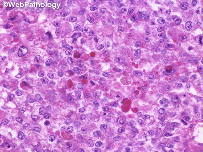
Comments:
Hepatoid pattern in Yolk Sac Tumor (seen in 20-40% of cases): The tumor cells have abundant eosinophilic cytoplasm, vesicular nuclei, and prominent nucleoli. They resemble fetal hepatocytes and are arranged in solid sheets, nests or trabeculae separated by fibrous septa. The morphology can mimic hepatocellular carcinoma, fibrolamellar type. They contain hyaline globules and are immunoreactive for AFP, alpha-1-antitrypsin, HepPar-1 and polyclonal CEA (canalicular pattern). Bile canaliculi-like structures are common; however, bile production is seen only rarely.
slide 115 of 183