
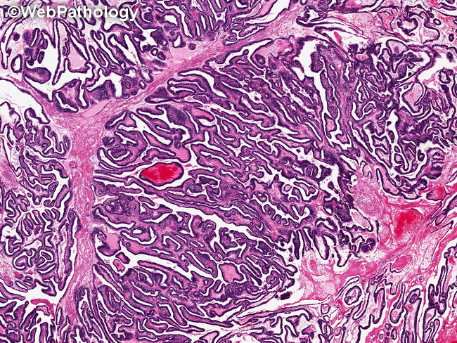
Yolk Sac Tumor : Glandular Pattern


Comments:
Glandular pattern in Yolk Sac Tumor (one-third of cases): Columnar tumor cells form simple tubules with round/oval lumen or complex anastomosing glandular structures. The pattern may be focal or more widespread. The glands may merge with the vesicles of polyvesicular vitelline pattern or may be present in a myxomatous, microcystic, or solid background. The glands may be primitive-appearing or well-differentiated and may resemble intestinal epithelium with Paneth cells and goblet cells. Some cases show subnuclear vacuoles similar to those seen in secretory endometrium. In rare cases (such as the one shown here), there is striking resemblance to endometrioid carcinoma. One must keep in mind that in older women, yolk sac tumor may be associated with endometrioid or clear cell carcinoma. Pure glandular yolk sac tumor is more common after chemotherapy and is therefore seen more frequently in metastases and late recurrences.