
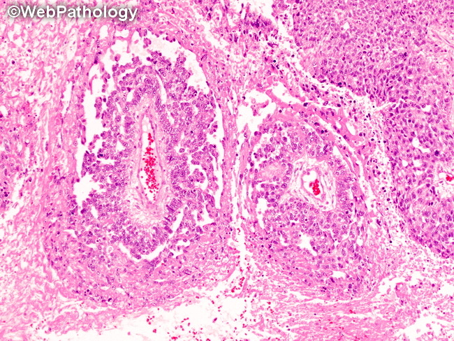
Yolk Sac Tumor : Schiller-Duval Body

slide 108 of 183


Comments:
Endodermal sinus (perivascular) pattern: This pattern displays papillary structures called Schiller-Duval body. It consists of a fibrovascular core with a single central thin-walled vessel in a hyalinized stroma surrounded by a one or more layers of cuboidal or columnar tumor cells with clear cytoplasm. The papillary structure is contained within a cystic space lined by flattened cells. Schiller-Duval body is a hallmark of yolk sac tumor but is not required for diagnosis and may not be seen in every case. Two large Schiller-Duval bodies are seen in this image.
slide 108 of 183