
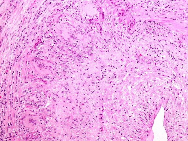
Giant Cell Arteritis : Microscopic Features

slide 10 of 17


Comments:
Microscopic Features: The normal architecture of the arterial wall is partially destroyed by a transmural lymphohistiocytic infiltrate. Multinucleated giant cells, which may contain phagocytized elastin debris, are usually present adjacent to the disrupted internal elastic lamina. The histologic changes may be segmental and a negative biopsy does not rule out the diagnosis. According to one study, only 60% of patients with clinical presentation of temporal arteritis have a positive biopsy. The specimen should be entirely submitted and subjected to serial sectioning. Image courtesy of: @PatholWalker
slide 10 of 17