
Cardiac Sarcoidosis : Gross Pathology


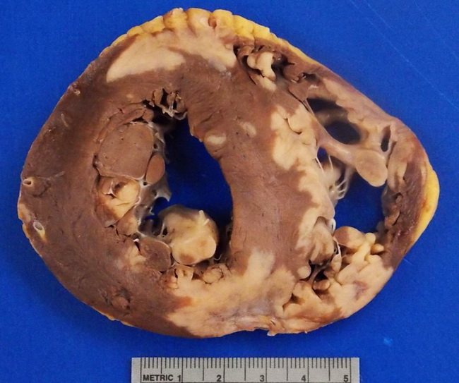
Comments:
Sarcoid granulomas may involve any part of the heart, including endocardium, myocardium, and pericardium. The most common sites are left ventricular free wall, papillary muscles, and basal ventricular septum. The granulomas are often distributed along the lymphatics which are especially numerous around cardiac conduction system tracts. This explains the pathogenesis of conduction blocks and ventricular arrhythmias (which are often fatal) in cardiac sarcoidosis. Grossly, the sarcoid lesions appear as diffuse, irregular, yellow-gray tumor-like infiltrates throughout the heart. Areas of fibrosis may be seen as slightly depressed gray-white scars. Abnormal thinning of ventricular wall and/or aneurysms may be present. This formalin-fixed slice of heart is from the same case as the previous two images. This 30 y/o male died suddently unexpectedly due to cardiac sarcoidosis. Image courtesy of: Megan Quinn, MD; used with permission.