
Renal Angiosarcoma : Pathology


Comments:
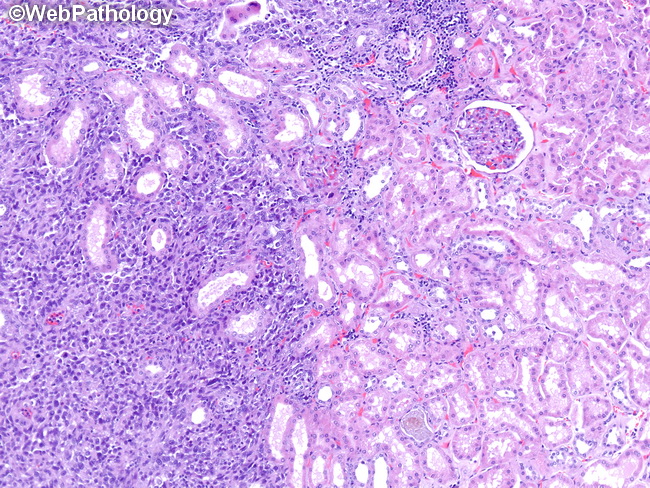
Gross and microscopic features of primary renal angiosarcomas are similar to those seen at other sites. Grossly, they consist of a single dominant mass with well-defined contours or multiple, ill-defined, hemorrhagic, spongy areas scattered throughout the kidney. Microscopically, well-differentiated tumors show anastomosing vascular channels lined by plump endothelial cells with variable degrees of cytologic atypia, hyperchromatic nuclei, and increased mitotic activity. Multilayering of endothelial cells with formation of papillary tufts is common. High-grade areas are more solid with spindle and/or epithelioid morphology. Like other sites, most renal angiosarcomas have an admixture of low- and high-grade areas. This image shows a solid focus of high-grade angiosarcoma infiltrating the renal parenchyma. Such cases would need a panel of immunohistochemical markers to differentiate them from various subtypes of renal cell carcinoma as well as metastases.