
Hepatic Angiosarcoma

slide 90 of 115


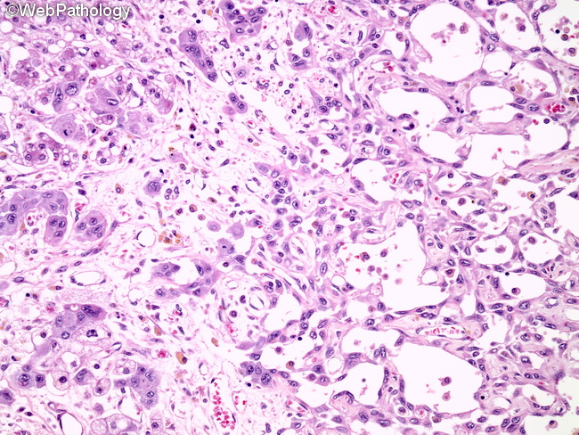
Comments:
This is another example of well-differentiated angiosarcoma of liver. The right half of the image shows anastomosing vascular channels lined by atypical endothelium with plump nuclei. The right half shows liver parenchyma with hepatocyte atrophy, fibrosis, and bile stasis.
slide 90 of 115