
Lymphedema-associated Angiosarcoma

slide 33 of 115


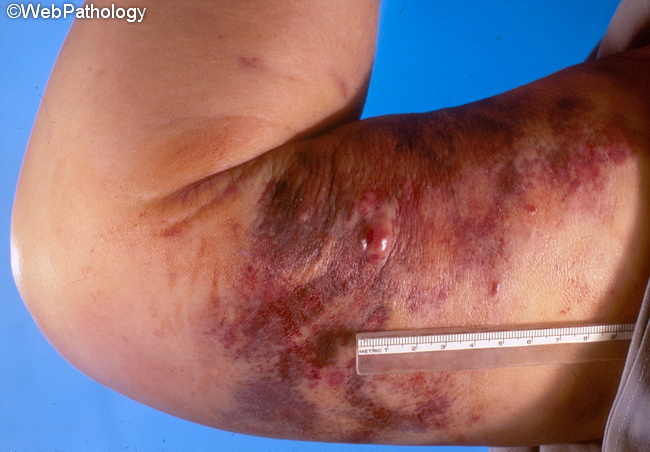
Comments:
Chronic lymphedema of any origin is one of the most common risk factors for the development of angiosarcoma of skin and soft tissues. Lymphedema-associated angiosarcoma has been encountered in the following locations and clinical settings:
- Arms of elderly females who had modified radical mastectomy with axillary node dissection for breast cancer (Stewart-Treves Syndrome)
- Abdominal wall following lymph node dissection for carcinoma of penis
- Congenital, idiopathic, iatrogenic, traumatic, or filarial lymphedema affecting an extremity
- Localized massive lymphedema related to morbid obesity.
slide 33 of 115