
Botryoid Rhabdomyosarcoma : Microscopic

slide 36 of 94


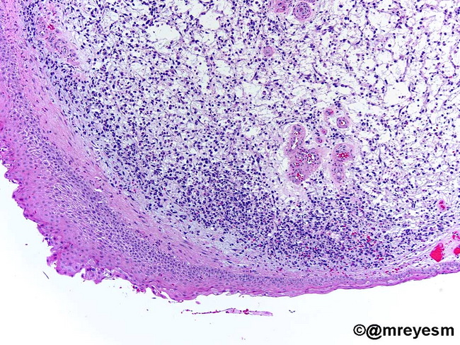
Comments:
Microscopic Features of Botryoid Rhabdomyosarcoma (RMS): The tumor cells range from primitive small cells with hyperchromatic nuclei to stellate cells with delicate cytoplasmic processes to spindle cells with rhabdomyoblastic differentiation. The background stroma is loose myxomatous type. The tumor cells form a zone that is several layers thick (including the cambium layer described earlier). The surface epithelium may show pseudoepitheliomatous hyperplasia mimicking carcinoma. There is strong immunoreactivity for myogenic antigens, including myogenin, muscle-specific actin, and desmin. Image courtesy of: Dr. Miguel Reyes-Mugica, Chief of Pediatric Pathology, University of Pittsburgh, PA, USA; used with permission.
slide 36 of 94